


| Product Name | CD38 Antibody |
|---|---|
| Antibody Type | Primary Antibodies |
| Antigen Alias | ADP-ribosyl cyclase/cyclic ADP-ribose hydrolase 1, 2'-phospho-ADP-ribosyl cyclase, 2'-phospho-ADP-ribosyl cyclase/2'-phospho-cyclic-ADP-ribose transferase, 2'-phospho-cyclic-ADP-ribose transferase, ADP-ribosyl cyclase 1, ADPRC 1, Cyclic ADP-ribose hydrolas |
| Product description | CD38 is a novel multifunctional ectoenzyme widely expressed in cells and tissues especially in leukocytes. CD38 also functions in cell adhesion,signal transduction and calcium signaling.1) Horenstein,A.L., Mol. Med. 15 (3-4), 76-84 (2009) Liu,Q., Biochemistry 47 (52), 13966-13973 (2008) Liu,Q., Chem. Biol. 15 (10), 1068-1078 (2008) |
| Immunogen | This CD38 antibody is generated from rabbits immunized with a KLH conjugated synthetic peptide between 241-270 amino acids from the C-terminal region of human CD38. |
| Clonality | Polyclonal |
|---|---|
| Isotype | Ig |
| Host Species | Rabbit |
| Tested Applications | FACSIFIHC-PWB |
| For WB starting dilution is: 1:1000 For IHC-P starting dilution is: 1:50~100 For IF starting dilution is: 1:10~50 For FACS starting dilution is: 1:10~50: |
|
| Species Reactivity | Human |
| Concentration | 1mg/ml |
| Gene Symbol | CD38 |
|---|---|
| Alternative Names | ADP-ribosyl cyclase/cyclic ADP-ribose hydrolase 1 2'-phospho-ADP-ribosyl cyclase 2'-phospho-ADP-ribosyl cyclase/2'-phospho-cyclic-ADP-ribose transferase 2'-phospho-cyclic-ADP-ribose transferase ADP-ribosyl cyclase 1 ADPRC 1 Cyclic ADP-ribose hydrolas |
| Molecular Weight(MW) | 34 kDa |
| Sequence Similarities | Predicted species reactivity based on immunogen sequence: Monkey |
Application
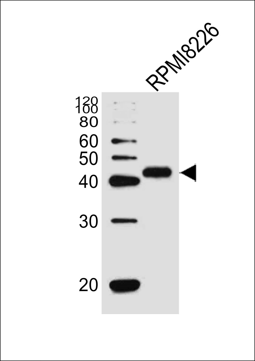
Western blot analysis of lysate from RPMI 8226 cell line, using CD38 Antibody at 1:1000.
Application
Confocal immunofluorescent analysis of CD38 Antibody with Hela cell followed by Alexa Fluor 488-conjugated goat anti-rabbit lgG (green). DAPI was used to stain the cell nuclear (blue).
Application
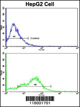
Flow cytometric analysis of HepG2 cells using CD38 Antibody (bottom histogram) compared to a negative control cell (top histogram). FITC-conjugated goat-anti-rabbit secondary antibodies were used for the analysis.
Application
Immunofluorescence analysis of CD38 Antibody with paraffin-embedded human hepatocarcinoma tissue . 0.05 mg/ml primary antibody was followed by FITC-conjugated goat anti-rabbit lgG (whole molecule). FITC emits green fluorescence.Red counterstaining is PI.
Application
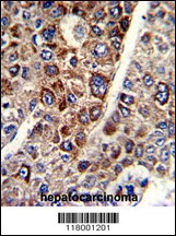
Formalin-fixed and paraffin-embedded human hepatocarcinoma with CD38 Antibody , which was peroxidase-conjugated to the secondary antibody, followed by DAB staining.
Application
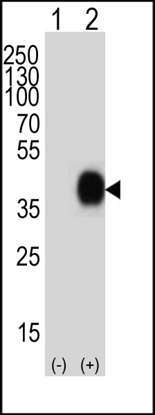
Western blot analysis of lysate from 293T cell line, using CD38 Antibody at 1:1000.
Application
Western blot analysis of lysate from RPMI8226 cell line, using CD38 Antibody at 1:1000.| Application Notes | For WB starting dilution is: 1:1000 For IHC-P starting dilution is: 1:50~100 For IF starting dilution is: 1:10~50 For FACS starting dilution is: 1:10~50: |
|---|
| Form | Liquid |
|---|---|
| Storage Instructions | Store at 4˚C for three months and -20˚C, stable for up to one year. As with all antibodies care should be taken to avoid repeated freeze thaw cycles. Antibodies should not be exposed to prolonged high temperatures. |
| Storage Buffer | Supplied in PBS with 0.09% (W/V) sodium azide. |


 Data sheet for OM274487